 This website is best viewed
using the horizontal display on
your tablet device.
This site is intended for Canadian
Healthcare Professionals only.
Healthcare Professionals only.
ENGLISH
FRANÇAIS
Stages of erythropoiesis
Erythropoiesis is a finely regulated, continuous process that can be conceptually divided into an early and late stage. Early-stage erythropoiesis is characterized by the proliferation of early-stage erythroid cells (progenitors), whereas late-stage erythropoiesis involves the differentiation and maturation of late-stage erythroid cells (precursors).1-3
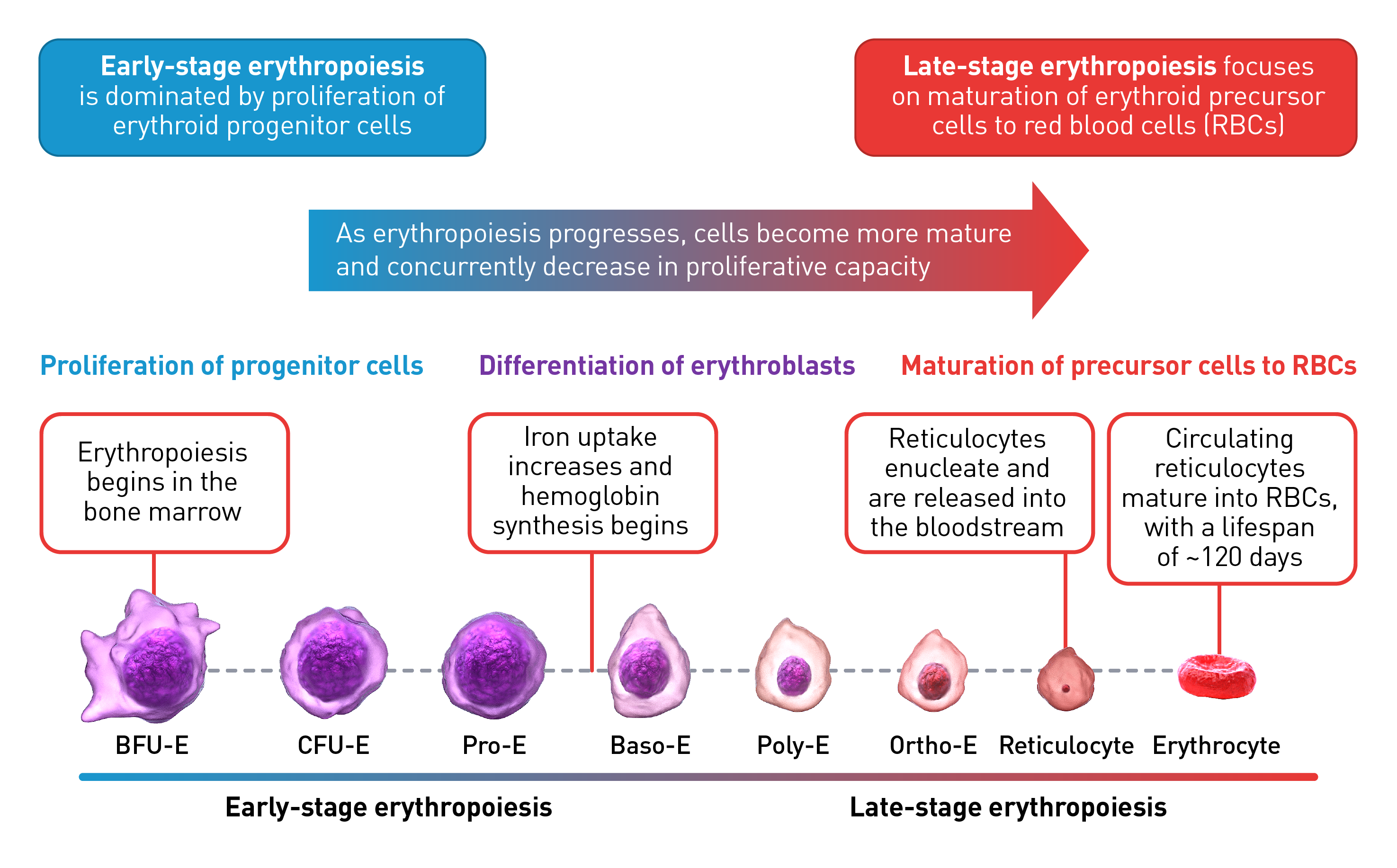
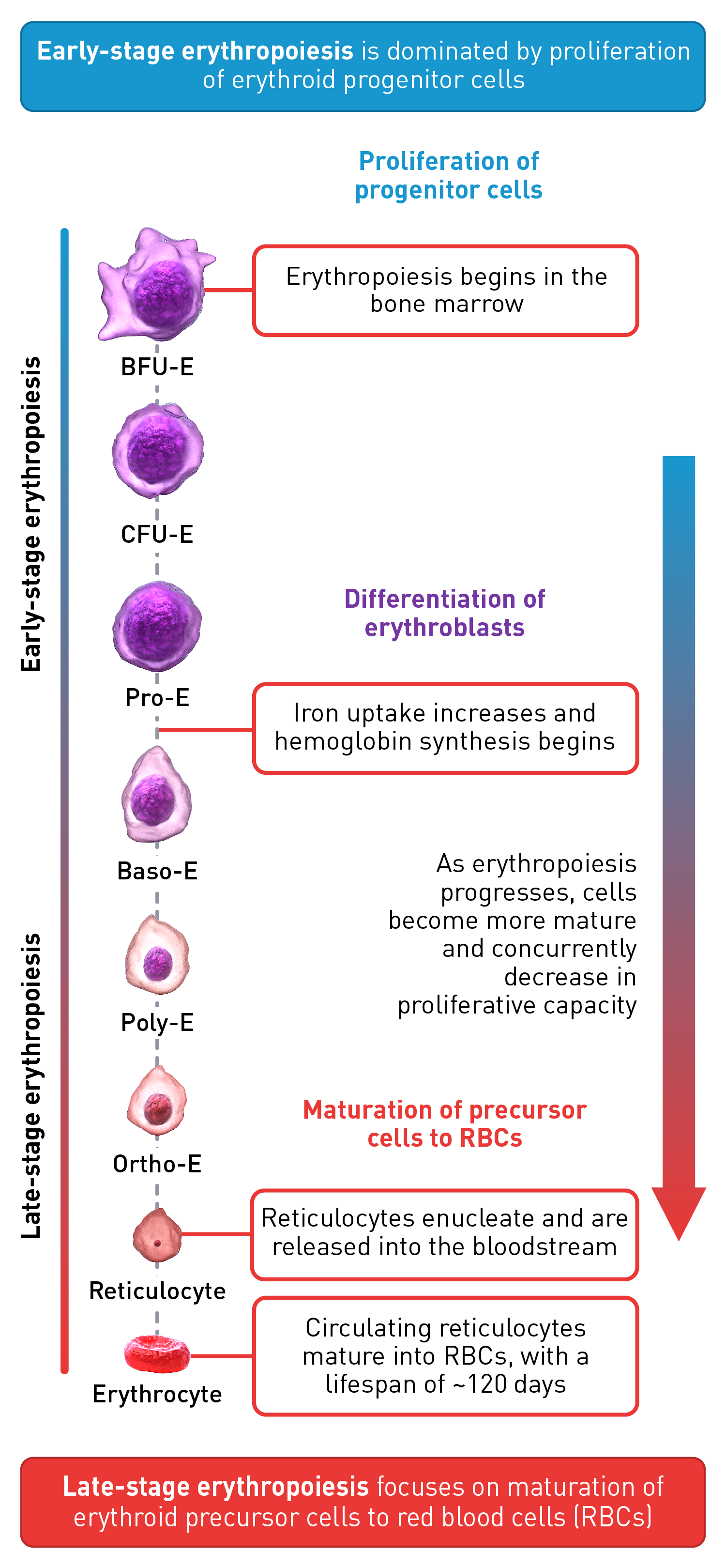
During the early stage of erythropoiesis, the proliferation of hematopoietic stem cells (HSCs) result in early erythroid progenitor cells. BFU-E represent the earliest progenitors committed exclusively to erythroid maturation that later differentiate into late CFU-E and Pro-E.3 Pro‑E, the earliest recognizable erythroid cells, enter terminal erythropoiesis and undergo morphological changes, such as in protein production and reduction in cell size and proliferative capacity. These changes lead to Pro-E differentiation and give rise to Baso‑E, Poly‑E and Ortho‑E, successively. At the end of this phase, erythroblasts expel their nuclei and lose all their organelles to produce mature enucleated cells, called reticulocytes.4-5 Once the nucleus is expelled, the reticulocyte is released into the bloodstream for maturation to continue and to produce fully functional, biconcave erythrocytes within 1–2 days.4
The late-stage maturation of red blood cells is an important step of erythropoiesis as it generates functional erythrocytes. Defects in this stage, known as erythroid maturation defects (EMDs), may impair the maturation process and result in ineffective erythropoiesis.6
The regulation of erythropoiesis
A complex network of transcription factors and epigenetic regulators are involved in regulating erythropoiesis. This regulatory network may adjust to changing pathological and physiological conditions. However, some pathological conditions may overwhelm the network, resulting in polycythemia or anemia.2
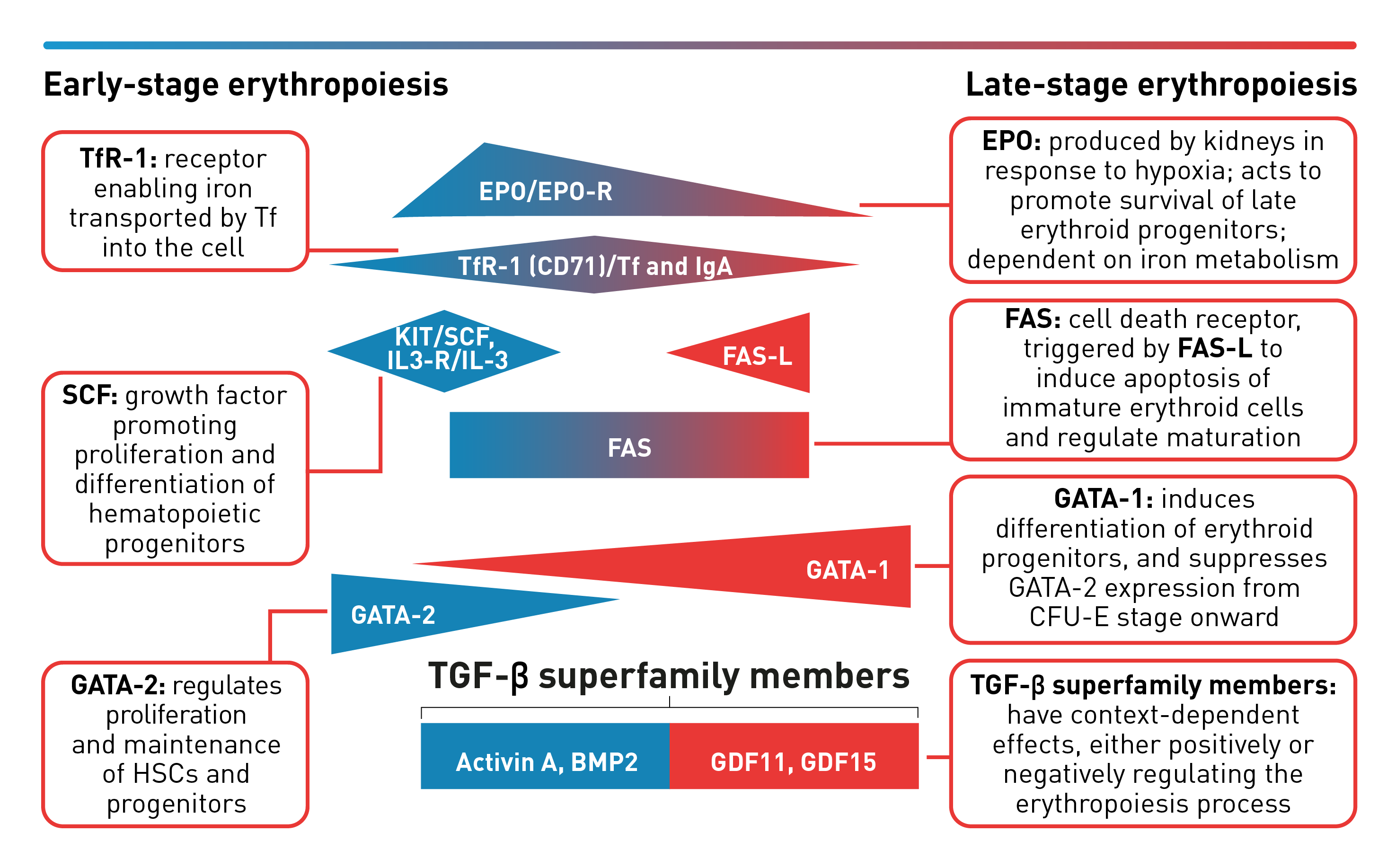
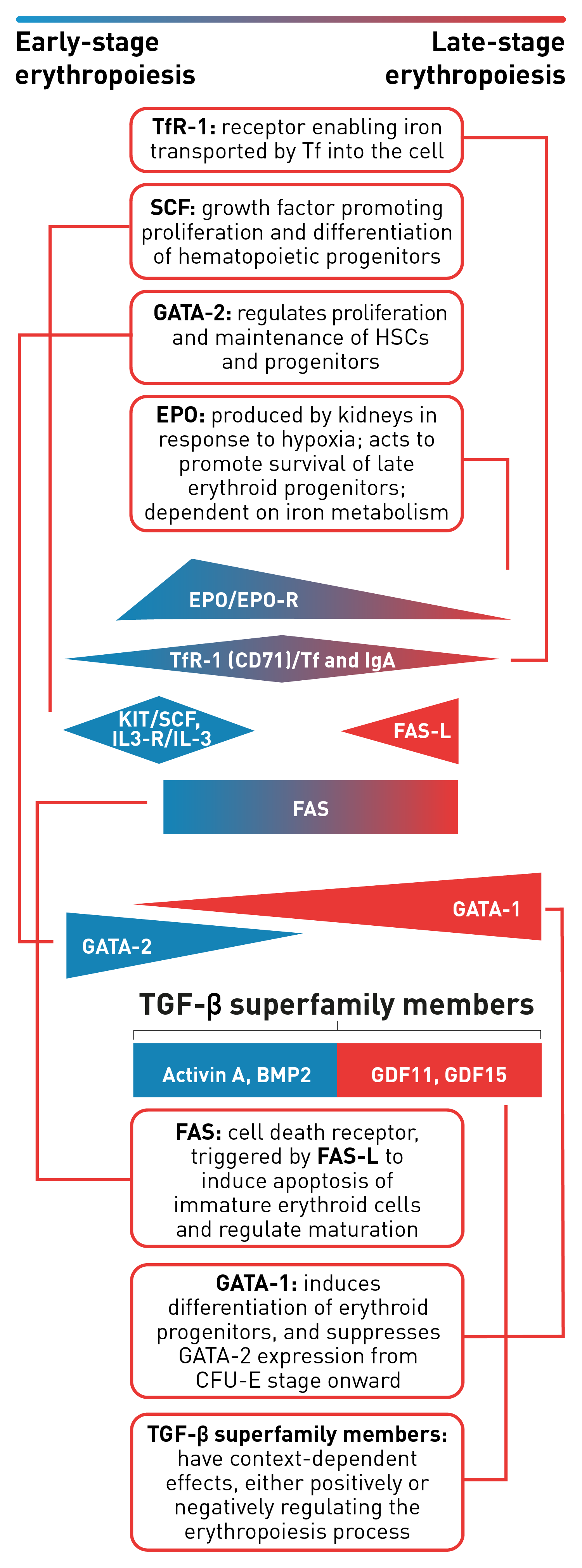
Erythropoiesis is tightly regulated to ensure it continues as a steady-state process. EPO, the main regulator of erythropoiesis, is responsible for promoting the survival and proliferation of erythroid progenitor cells by preventing apoptosis.3 GATA‑1 is the main regulator of lineage-commitment, differentiation and survival of early-stage erythroid cells. Other transcription factors include TfR1, an iron metabolism regulator, and SCF, an early acting hematopoietic growth factor. FAS‑L is expressed by late-stage erythroid cells to trigger apoptosis of immature erythroid cells and regulate maturation, while selected TGF‑β superfamily ligands play a key role in regulating erythroid maturation.2
Transcription factors and regulators involved in erythropoiesis
Erythropoietin (EPO)
EPO is a key positive regulator of early-stage erythropoiesis that helps to stimulate red blood cell production. It mainly acts on CFU-E, Pro-E and early-stage Baso-E, before it gradually wanes in later maturation stages.2,7
GATA-1 and GATA-2
The transcription factor, GATA‑2, is highly expressed in HSCs and in early-stage progenitors, but its expression is suppressed in the CFU‑E stage and onward. During the CFU-E stage, the expression of GATA-1 in CFU‑E progenitors is activated and increases as the erythroblasts differentiate and mature. The transition of GATA expression, from GATA‑2 to GATA‑1, is referred to as the GATA switch mechanism. This mechanism results in the mutually exclusive expression of GATA‑2 and GATA‑1 during early and late stages of erythropoiesis, respectively.8
FAS ligand (FAS-L)
FAS-L is expressed by late-stage erythroid precursor cells and is involved in regulating the final maturation stages that lead to the generation of functional red blood cells.2
Transforming growth factor β (TGF-β) superfamily
Members of the TGF‑β superfamily can exert opposing regulatory effects on the erythropoiesis process.9,10,11 Signalling mediated by selected TGF‑β superfamily ligands helps to regulate erythroid maturation through the SMAD pathway.11 Dysregulation of this signalling pathway may contribute to impaired erythroid maturation, leading to ineffective erythropoiesis.9,10
Expert discussion: Mechanisms of erythropoiesis and erythroid homeostasis

Professor Olivier Hermine, Professor of Haematology, Paris Necker Children’s Hospital
In this video, Professor Hermine outlines the mechanism of erythropoiesis and explains the importance of erythroid homeostasis, highlighting the regulatory hormones and transcription factors involved.
 3-minute video
Baso-E: Basophilic erythroid, BFU-E: Burst-forming unit-erythroid, CFU-E: Colony-forming unit-erythroid, Ortho-E: Orthochromatophilic, Poly-E: Polychromatophilic, Pro-E: Proerythroblasts, BMP2: Bone morphogenetic protein 2, EPO: Erythropoietin, EPO-R: Erythropoietin receptor, FAS-L: FAS ligand, GDF: Growth differentiation factor, IL-3: Interleukin 3, IL-3-R: Interleukin 3 receptor, IgA: Immunoglobulin A, SCF: Stem cell factor, Tf: Transferrin, TfR-1 (or CD71): Transferrin receptor, TGF: Transforming growth factor.
 This website is best viewed
using the vertical display on
your mobile device.